


Prostate cancer is one of the most common malignancies in aging men, requiring a structured clinical and histological diagnostic evaluation to differentiate it from prevalent benign urological conditions that present with overlapping signs, biological markers, and imaging characteristics.
1. Clinical Overview and Diagnostic Rationale
What is Prostate Cancer?
Prostate cancer is defined as an invasive malignant neoplasm originating from the glandular epithelial cells of the prostate gland, predominantly developing within the peripheral zone as adenocarcinoma. It develops through multi-step genetic and epigenetic mutations that compromise cell cycle regulation, leading to unchecked cellular proliferation, tissue invasion, and metastasis.
Why Differential Diagnosis Matters
Diagnosing prostate cancer accurately is challenging because its early manifestations closely mimic several non-malignant lower urinary tract disorders. Conditions such as benign prostatic hyperplasia (BPH), acute or chronic bacterial prostatitis, prostatic abscesses, and urinary tract stone disease can cause similar lower urinary tract symptoms (LUTS), elevate serum prostate-specific antigen (PSA) levels, and produce abnormal findings on digital rectal examinations (DRE) or transrectal ultrasonography (TRUS).

2. Primary Urological Conditions Confounded with Prostate Cancer
1. Benign Prostatic Hyperplasia (BPH)
- Pathophysiological Overlap: BPH involves non-malignant nodular hyperplasia of both stromal and epithelial elements within the transition zone of the prostate gland, driven by aging and dihydrotestosterone (DHT) stimulation. Like prostate cancer, BPH causes urinary frequency, urgency, nocturia, weak stream, and hesitancy.
- Diagnostic Differentiation: While BPH primarily expands inward to compress the prostatic urethra from the transition zone, prostate cancer predominantly originates in the peripheral zone. On DRE, BPH presents as a smooth, symmetrically enlarged, firm or elastic prostate, whereas malignancy manifests as indurated, hard, asymmetrical nodules. Serum PSA levels can be elevated in BPH due to increased prostatic volume, but calculating PSA density (PSA /prostate volume) and evaluating free-to-total PSA ratios help differentiate benign growth from malignancy.
2. Acute and Chronic Prostatitis
- Pathophysiological Overlap: Prostatic inflammation damages glandular epithelial cells, causing significant leakage of intracellular tissue kallikrein (PSA) into the systemic circulation. This results in transient, marked elevations in serum total PSA that can easily be misdiagnosed as prostate cancer.
- Diagnostic Differentiation: Acute bacterial prostatitis presents with acute systemic signs, including high fever, chills, severe dysuria, and perineal pain. On DRE, the prostate is exquisitely tender, warm, and boggy; DRE must be performed with extreme caution to prevent bacteremia. Serum PSA levels elevated during acute prostatitis normalize or decrease significantly 4 to 8 weeks after appropriate antibiotic therapy, whereas PSA elevations in prostate cancer remain persistently high or continue to rise.
3. Prostatic Abscess and Granulomatous Prostatitis
- Pathophysiological Overlap: Prostatic abscesses or severe chronic granulomatous prostatitis (secondary to BCG therapy, tuberculosis, or idiopathic autoimmune reactions) form focal indurated areas that mimic malignant tumors on digital rectal examination and TRUS imaging.
- Diagnostic Differentiation: Pelvic multiparametric MRI (mpMRI) reveals characteristic fluid collection with peripheral rim enhancement in prostatic abscesses. Granulomatous lesions require histopathological confirmation via core needle biopsy to rule out adenocarcinoma.
4. Prostatic Calculi and Urethral Stricture Disease
- Pathophysiological Overlap: Calcifications within the prostatic acini generate hard, focal nodules palpable on DRE, simulating peripheral zone carcinoma. Urethral strictures create severe urinary outflow obstruction that reproduces the voiding LUTS seen in locally advanced prostate cancer.
- Diagnostic Differentiation: Transrectal ultrasonography (TRUS) or non-contrast pelvic CT clearly demonstrates hyper-echoic prostatic calculi with posterior acoustic shadowing. Urethral strictures are definitively diagnosed using retrograde urethrography (RUG) or flexible cystoscopy.
3. Comparative Clinical and Diagnostic Matrix
| Urological Condition | Dominant Anatomical Zone | Digital Rectal Exam (DRE) Findings | PSA Profile Characteristics | Key Distinguishing Diagnostic Tool |
| Prostate Cancer | Peripheral Zone (70%-75%) | Hard, asymmetric nodule, loss of sulcus | Persistent elevation, low %Free PSA (<15%) | mpMRI (PI-RADS 4-5), Core Needle Biopsy |
| Benign Prostatic Hyperplasia | Transition Zone | Symmetrically enlarged, smooth, elastic | Normal to mildly elevated, normal %Free PSA | Normal mpMRI (PI-RADS 1-2), TRUS volume calculation |
| Acute Prostatitis | Diffuse / Global Gland | Exquisitely tender, hot, boggy gland | Marked transient elevation (>20-50 ng/mL) | Urinalysis, urine culture, post-antibiotic PSA decline |
| Prostatic Abscess | Focal Parenchyma | Fluctuant, tender focal area | Variable, elevated during acute flare | Pelvic Ultrasound / MRI showing rim-enhancing cavity |
| Prostatic Calculi | Acinar Ducts | Hard, focal nodular sensation | Typically normal unless co-existing BPH/infection | TRUS showing hyper-echoic foci with acoustic shadowing |
4. Comprehensive Diagnostic Pathways for Prostate Cancer
Establishing an accurate diagnosis requires a structured pathway moving from non-invasive screening to definitive histopathology.

- Digital Rectal Examination (DRE): Evaluates prostate size, contour, symmetry, and detects suspicious indurated nodules in the peripheral zone.
- Serum Prostate-Specific Antigen (PSA) Assay: Measures total serum PSA. Free-to-total PSA ratios (%Free PSA ) below 15% significantly increase the probability of prostate cancer, whereas higher ratios point toward BPH.
- Multiparametric Pelvic MRI (mpMRI): The non-invasive gold standard imaging technique combining T2-weighted imaging, Diffusion-Weighted Imaging (DWI), and Dynamic Contrast Enhancement (DCE). Findings are reported using the PI-RADS system (PI-RADS 1 to 5).
- Prostate Core Needle Biopsy: Definitive diagnosis relies on histopathological confirmation obtained via TRUS-guided or transperineal MRI-fusion targeted core needle biopsy. Malignant tissues are classified using the Gleason Grading System and ISUP Grade Groups (Groups 1 through 5).
5. Medical Therapy and Pharmacological Principles
Pharmacotherapy plays a central role in managing advanced or metastatic prostate cancer and co-existing benign conditions.
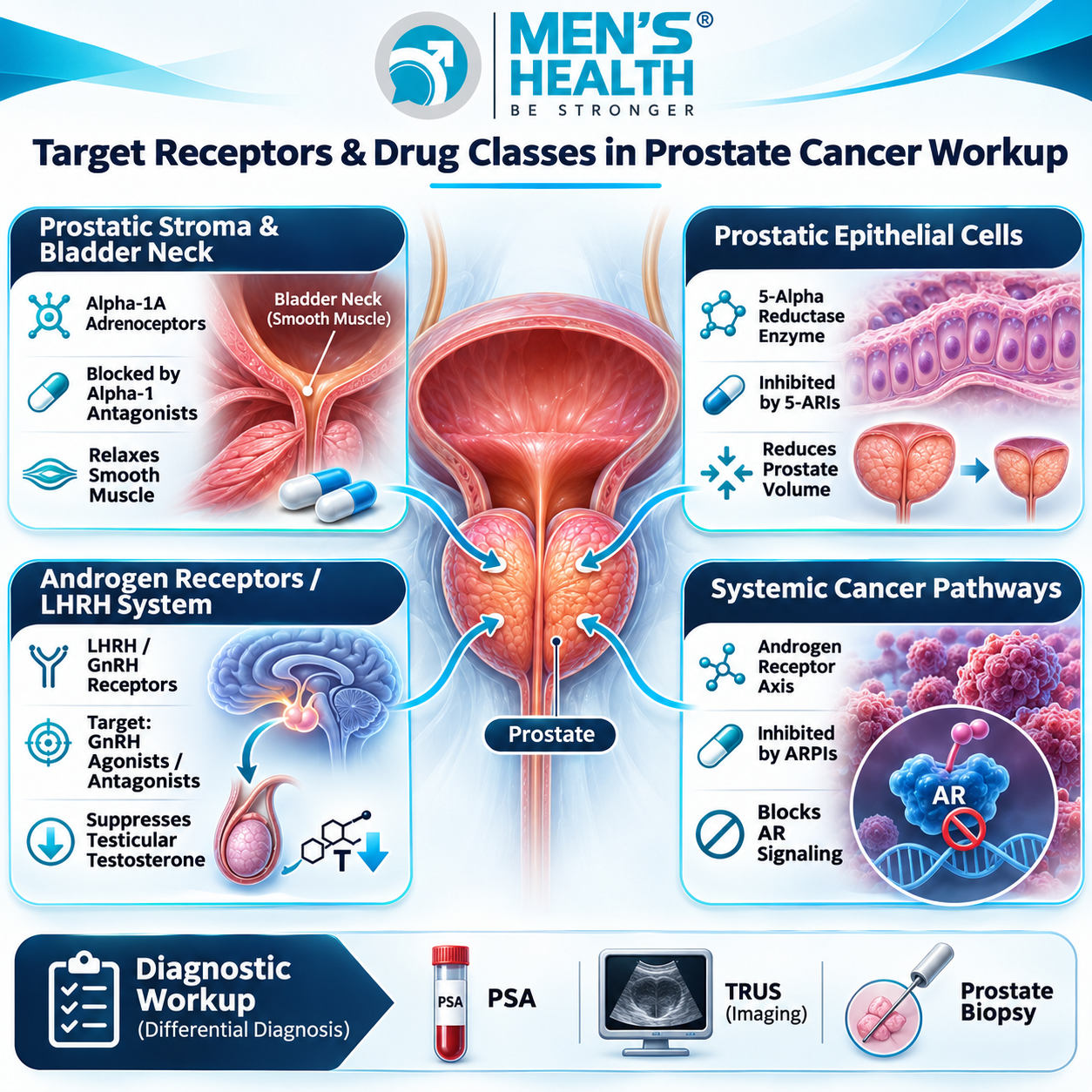
Core Pharmacological Classes
Androgen Deprivation Therapy (ADT – GnRH Agonists & Antagonists)
- Mechanism of Action: Suppresses testicular testosterone production. GnRH agonists (e.g., Leuprolide, Goserelin) initially cause a transient testosterone flare before downregulating pituitary receptors, whereas GnRH antagonists (e.g., Degarelix, Relugolix) achieve immediate testosterone suppression without a surge.
- Clinical Indications: Advanced, metastatic, or high-risk localized prostate cancer combined with radiotherapy.
- Precautions & Adverse Effects: Hot flashes, loss of bone mineral density (osteoporosis), sarcopenia, metabolic syndrome, fatigue, and cardiovascular risk.
Novel Androgen Receptor Pathway Inhibitors (ARPIs)
- Mechanism of Action: Block androgen binding to androgen receptors, inhibit nuclear translocation, and prevent DNA binding.
- Representative Agents: Enzalutamide, Abiraterone acetate, Apalutamide, Darolutamide.
- Clinical Indications: Metastatic hormone-sensitive, non-metastatic castration-resistant, and metastatic castration-resistant prostate cancer.
- Precautions: Requires monitoring of hypertension, hepatic transaminases, fatigue, and drug-drug interactions via CYP450 pathways.
Medications for Co-Existing Benign Conditions (α1-Blockers & 5-ARIs)
- α1-Adrenoceptor Antagonists (e.g., Tamsulosin, Silodosin): Prescribed to manage lower urinary tract symptoms secondary to BPH.
- 5-α Reductase Inhibitors (5-ARIs – e.g., Finasteride, Dutasteride): Inhibits conversion of testosterone to DHT, shrinking prostatic volume in BPH. Clinical Guidance: 5-ARIs lower baseline total PSA by approximately 50% after 6 months of continuous therapy; failure of PSA to decline by half should prompt investigation for occult prostate cancer.
6. Comprehensive Care at Men’s Health Center and Men’s Health Pharmacy
Diagnostic evaluation and long-term management of prostate cancer and overlapping urological conditions require coordinated medical and pharmaceutical support.

Men’s Health Pharmacy, an integral component of the Men’s Health Center ecosystem founded by Dr. Tra Anh Duy (MD, PhD, Spec. II), provides clinical pharmacy support for male urological health. At Men’s Health, patients consult with experienced urologists and clinical pharmacists to ensure that prescribed therapeutic regimens – including ADT agents, ARPIs, α1-blockers, and 5-ARIs – are selected safely, screened for drug interactions, and monitored effectively. The facility emphasizes professional safety, patient privacy, absolute confidentiality, and evidence-based clinical practice across all stages of care.
7. Lifestyle Modifications and Preventive Strategies
- Balanced Nutrition: Adopt a Mediterranean-style diet rich in vegetables, legumes, omega-3 fatty acids, and antioxidants while reducing processed foods and saturated fats.
- Regular Physical Activity: Engaging in 150 minutes of moderate aerobic and resistance exercise weekly maintains bone density and counters metabolic side effects during androgen deprivation therapy.
- Weight Management: Maintaining a healthy body mass index (BMI) reduces systemic low-grade inflammation associated with aggressive prostate cancer biology.
- Routine Urological Screening: Men aged 50 and older (or age 40–45 for those with a family history of prostate cancer or BRCA mutations) should discuss baseline PSA testing and clinical risk evaluation with a urologist.
8. Common Clinical Pitfalls in Differential Diagnosis
- Assuming Elevated PSA Always Means Cancer: Performing invasive biopsies without evaluating for active urinary tract infections or acute prostatitis, leading to unnecessary procedures.
- Ignoring the Effect of 5-ARIs on Serum PSA: Failing to double the measured PSA value in patients on long-term finasteride or dutasteride therapy, resulting in missed prostate cancer diagnoses.
- Overlooking Digital Rectal Examination: Relying solely on PSA screening without performing DRE, potentially missing PSA-negative high-grade prostate malignancies.
- Delaying mpMRI Prior to Biopsy: Performing systematic blind biopsies without pre-biopsy pelvic mpMRI increases the risk of under-sampling clinically significant tumors.
9. Frequently Asked Questions
Can Benign Prostatic Hyperplasia (BPH) turn into prostate cancer?
No. BPH and prostate cancer are distinct pathological entities. BPH involves benign cellular hyperplasia primarily in the transition zone, whereas prostate cancer develops as malignant cellular transformation mainly in the peripheral zone. However, both conditions can coexist in aging men.
Why does acute prostatitis elevate PSA levels so dramatically?
Acute prostatitis causes severe inflammation and acinar cell damage within the prostate gland. This disruption causes intracellular PSA to leak directly into the systemic bloodstream, elevating total serum PSA levels transiently.
What is the role of multiparametric MRI (mpMRI) in differentiating prostate cancer from benign conditions?
Pelvic mpMRI combines anatomical (T2-weighted) and functional (DWI, DCE) imaging sequences to assign a PI-RADS score (1 to 5). It accurately highlights clinically significant suspicious malignant lesions while helping rule out benign hyperplasia or inflammation.
How does finasteride or dutasteride affect prostate cancer screening?
5-α reductase inhibitors reduce prostate volume and cut measured serum total PSA levels approximately in half after 6 months of continuous use. Clinicians must double the measured PSA value in these patients to maintain accurate screening thresholds.
When should a man undergo a prostate biopsy?
A prostate biopsy is indicated when a patient presents with a persistently elevated serum PSA level, an abnormal free-to-total PSA ratio, a suspicious lesion on pelvic mpMRI (PI-RADS 4 or 5), or an abnormal indurated nodule on Digital Rectal Examination.
Conclusion and Expert Clinical Advice
Accurately diagnosing prostate cancer requires careful clinical differentiation from common overlapping urological conditions like Benign Prostatic Hyperplasia, acute or chronic prostatitis, and prostatic abscesses. Employing a structured diagnostic framework – combining digital rectal examination, PSA kinetics, free-to-total PSA ratios, multiparametric pelvic MRI, and core needle biopsy – ensures accurate identification of malignancy while avoiding unnecessary procedures for benign conditions.
Patients experiencing lower urinary tract symptoms, urinary frequency, weak stream, or elevated PSA readings should avoid self-diagnosis or unprescribed medications. Seeking clinical evaluation at a specialized urological center such as the Men’s Health Center guarantees precise diagnostic assessment, specialist physician management, and dedicated clinical pharmacy support for long-term urological health.