
Urinary retention in men is a critical lower urinary tract disorder characterized by the inability to empty the bladder effectively, requiring targeted clinical, laboratory, and urodynamic assessments to differentiate acute emergency presentation from silent chronic pathology.
1. Clinical Overview and Diagnostic Rationale
Defining Acute vs. Chronic Urinary Retention
Urinary retention occurs when intravesical pressure generated during the voiding phase fails to overcome bladder outlet resistance, or when the detrusor muscle lacks sufficient contractility.
- Acute Urinary Retention (AUR): Defined as a sudden, complete inability to void voluntarily, accompanied by severe, agonizing suprapubic pain, rapid onset over hours, and a distended, exquisitely tender urinary bladder.
- Chronic Urinary Retention (CUR): Characterized as persistent, progressive incomplete bladder evacuation resulting in high post-void residual (PVR) volumes (typically ). CUR is frequently non-painful or indolent due to gradual bladder stretch adaptation, often presenting as urinary frequency, hesitancy, weak stream, or overflow (paradoxical) incontinence.
Pathophysiological Mechanisms
Micturition is regulated by a balanced neural cascade involving parasympathetic motor control ( and muscarinic receptors driving detrusor contraction), sympathetic storage regulation (-adrenoceptors mediating bladder neck/prostatic smooth muscle tone), and somatic innervation of the external urethral sphincter.
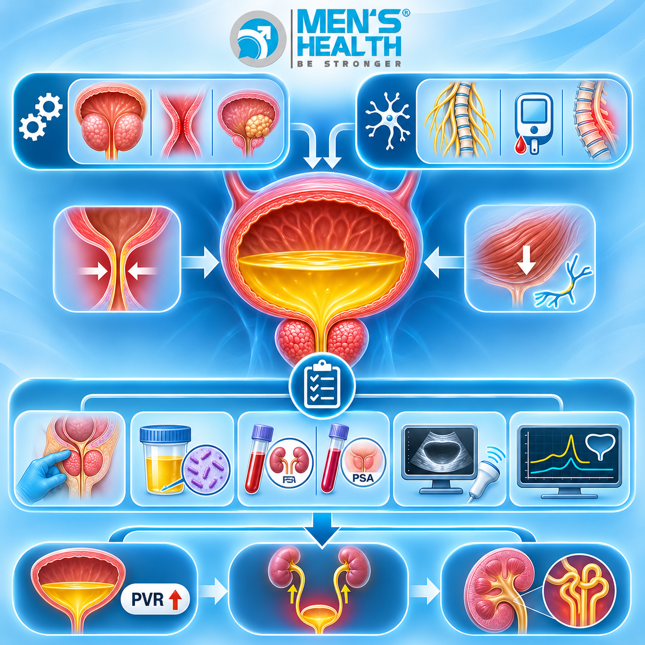
Urinary retention manifests via two main pathophysiological pathways:
- Mechanical Outflow Obstruction: Physical narrowing of the urethral lumen or bladder neck, primarily resulting from benign prostatic hyperplasia (BPH), prostate adenocarcinoma, urethral stricture disease, bladder neck contractures, or urethral stone impaction.
- Functional and Neurogenic Detrusor Failure: Diminished detrusor contractility (detrusor underactivity, DUA) caused by neurogenic lesions (stroke, Parkinson’s disease, spinal cord trauma, diabetic autonomic neuropathy) or myogenic degeneration secondary to chronic overdistension.
2. Primary Etiologies and Predisposing Risk Factors
Underlying Anatomical and Neurogenic Causes
- Benign Prostatic Hyperplasia (BPH): Adenomatous hypertrophy of the prostatic transition zone compresses the prostatic urethra, representing the leading mechanical etiology in men over 50 years of age.
- Urethral Stricture Disease: Fibrotic narrowing of the anterior or posterior urethral lumen following trauma, iatrogenic urethral catheterization injury, or prior gonococcal urethritis.
- Prostatic and Pelvic Malignancies: Advanced prostate cancer, bladder neck urothelial carcinoma, or extrinsic pelvic tumors compressing the lower tract.
- Acute Bacterial Prostatitis: Inflammatory edema within the prostatic stroma causing acute luminal compression.
- Neurogenic Bladder Dysfunction: Loss of cortical or sacral micturition center integration due to multiple sclerosis, spinal cord lesions, acute stroke, or severe diabetic neuropathy.
- Pharmacological Triggers: Systemic administration of medications possessing anticholinergic activity, first-generation -antihistamines, tricyclic antidepressants (TCAs), -adrenergic agonists (decongestants), or opioid analgesics.
Key Risk Factors
- Advanced age ( years).
- Pre-existing, untreated lower urinary tract symptoms (LUTS).
- Long-standing, poorly controlled Type 2 Diabetes Mellitus.
- History of pelvic radiotherapy, radical pelvic surgery, or lower tract instrumentation.
3. Clinical Manifestations and Red Flag Symptoms
Symptom Spectrum Matrix
| Clinical Feature | Acute Urinary Retention (AUR) | Chronic Urinary Retention (CUR) |
|---|---|---|
| Symptom Onset | Sudden (hours) | Insidious (weeks, months, or years) |
| Pain Level | Severe, excruciating suprapubic agony | Painless, mild discomfort, or dull pressure |
| Micturition Capability | Total inability to void | Ability to pass small, frequent amounts |
| Incontinence Pattern | Absent | Overflow (paradoxical) incontinence |
| PVR Volume | High (500−1000+ mL) | Markedly elevated (500−2000+ mL) |
| Primary Complication Risk | Bladder ischemia, extreme distress | Hydronephrosis, post-renal renal failure |
Red Flag Indications Requiring Immediate Medical Assessment
Immediate specialist evaluation is non-negotiable under the following clinical circumstances:
- Sudden, complete inability to void for hours associated with severe suprapubic pain.
- Systemic features of infection (fever, rigors, confusion) in a patient with bladder distension.
- Progressive neurological deficits (saddle anesthesia, lower extremity weakness, loss of anal sphincter tone).
- Gross hematuria or clot retention causing mechanical catheter or urethral occlusion.
- Uremic signs (persistent nausea, anorexia, vomiting, peripheral edema) indicative of post-renal kidney injury.
4. Comprehensive Diagnostic Pathways and Test Selection
Establishing the exact underlying cause of male urinary retention requires a structured diagnostic approach starting from clinical history to advanced urodynamics.
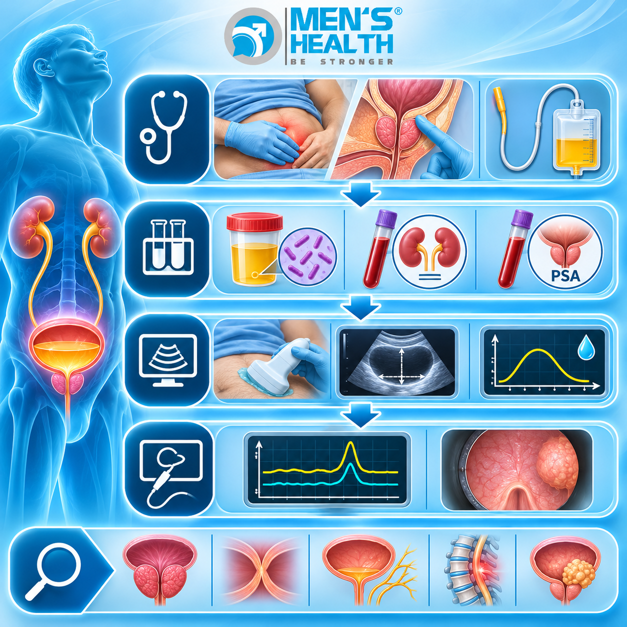
Essential Laboratory Diagnostic Tests
- Urinalysis and Dipstick Examination: Evaluates nitrites, leukocyte esterase, microscopic hematuria, and proteinuria to exclude acute urinary tract infection, pyuria, or underlying glomerular pathology.
- Urine Culture and Antibiotic Susceptibility: Essential to confirm bacterial cystitis or acute prostatitis, identifying specific pathogens and guiding anti-infective therapy.
- Serum Creatinine and Estimated Glomerular Filtration Rate (eGFR): Evaluates renal parenchyma function. Elevated creatinine indicates post-renal acute kidney injury (AKI) or chronic kidney disease (CKD) secondary to high-pressure back-pressure effects.
- Serum Electrolyte Panel (Sodium, Potassium, Bicarbonate): Mandatory in chronic retention decompressed via catheterization to monitor for electrolyte disturbances associated with post-obstructive diuresis.
- Serum Prostate-Specific Antigen (PSA): Measures prostatic glandular activity. Clinical Pearl: PSA testing should be deferred during an episode of acute retention or immediate catheter insertion, as acute distension and instrumentation cause transient PSA elevations leading to false-positive malignant suspicion. PSA should be re-evaluated 4 to 6 weeks after acute resolution.
Imaging and Functional Diagnostic Modalities
- Post-Void Residual (PVR) Ultrasound Scan: Point-of-care transabdominal ultrasound quantifying residual volume immediately post-micturition. A PVR >150−200 mL is abnormal; a PVR >300 mL is diagnostic of chronic urinary retention.
- Renal and Bladder Ultrasonography: High-resolution upper tract imaging to evaluate renal parenchymal thickness, screen for pelvicalyceal dilation, bilateral hydronephrosis, ureterectasia, and structural bladder wall abnormalities (e.g., detrusor trabeculation, diverticula, or mucosal calculi).
- Uroflowmetry: Non-invasive evaluation of urinary flow dynamics measuring maximum flow rate (Qmax), voided volume, and flow curve morphology. A maximum flow rate Qmax<10 mL/s with a voided volume >150 mL strongly indicates outflow obstruction or detrusor contractility failure.
- Multi-Channel Urodynamic Studies (UDS): Simultaneous measurement of intravesical pressure, intra-abdominal pressure, and flow rates during filling cystometry and pressure-flow voiding phases. UDS is the gold standard for differentiating Bladder Outlet Obstruction (BOO) from Detrusor Underactivity (DUA).
- Flexible Cystourethroscopy: Direct endoscopic visualization of the urethral passage, external sphincter, prostatic urethra, bladder neck, and mucosal bladder wall. It definitively confirms or excludes urethral strictures, bladder neck contractures, prostatic occlusive lobes, and intravesical neoplasms.
5. Diagnostic Summary Matrix for Male Urinary Retention
| Diagnostic Test | Primary Clinical Target | Key Diagnostic Findings |
|---|---|---|
| Urinalysis & Culture | Urinary tract infection & pyuria | Confirms presence of bacterial infection or microhematuria |
| Serum Creatinine & eGFR | Post-renal kidney function | Identifies acute kidney injury or chronic renal compromise |
| Post-Void Residual (PVR) | Residual intravesical volume | Quantifies residual urine (>300 mL confirms chronic retention) |
| Renal Ultrasonography | Upper urinary tract integrity | Identifies bilateral hydronephrosis, renal atrophy, or stones |
| Uroflowmetry | Micturition flow rate (Qmax) | Qmax <10 mL/s demonstrates impaired emptying dynamics |
| Urodynamic Studies (UDS) | Pressure-flow parameters | Distinguishes bladder outlet obstruction from detrusor underactivity |
| Flexible Cystoscopy | Lower tract structural luminal integrity | Visualizes urethral strictures, prostate obstruction, or bladder tumors |
6. Pharmacological Management Strategies
Once acute relief has been achieved or chronic diagnostics are completed, targeted medical therapy aims to lower outflow resistance, modify prostatic structural volume, or optimize detrusor function.
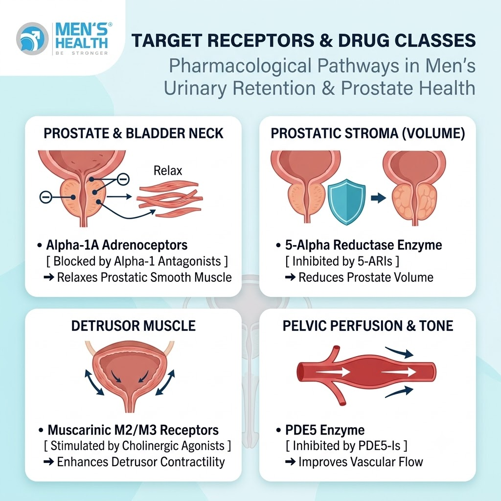
Main Pharmacological Classes
Alpha-1 Adrenoceptor Antagonists (-Blockers)
- Mechanism of Action: Selective antagonism of -adrenoceptors within the prostatic stroma, capsule, and bladder neck. Inhibits sympathetic smooth muscle tone, decreasing outflow resistance without altering detrusor contractility.
- Representative Agents: Tamsulosin, Silodosin, Alfuzosin, Doxazosin.
- Clinical Role: First-line pharmacotherapy prior to conducting a Trial Without Catheter (TWOC) following an acute retention episode, or for ongoing symptomatic BPH management.
- Adverse Effect Profile & Precautions: Orthostatic hypotension, dizziness, fatigue, nasal congestion, and retrograde ejaculation. Can induce Intraoperative Floppy Iris Syndrome (IFIS) during cataract extraction.
5-Alpha Reductase Inhibitors (5-ARIs)
- Mechanism of Action: Inhibit the -reductase enzyme (Types 1 and 2), blocking the conversion of testosterone into dihydrotestosterone (DHT). Reduces intraprostatic DHT levels, inducing apoptosis of epithelial cells and shrinking prostate volume by approximately over 3 to 6 months.
- Representative Agents: Finasteride, Dutasteride.
- Clinical Role: Indicated for long-term reduction of urinary retention risk and surgical need in men with documented prostate volume .
- Adverse Effect Profile & Precautions: Reduced libido, erectile dysfunction, ejaculatory failure, and gynecomastia. Patients must be counseled that serum PSA values are reduced by approximately after 6 months of continuous therapy.
Cholinergic Agonists (Parasympathomimetics)
- Mechanism of Action: Direct activation of muscarinic receptors on detrusor smooth muscle, increasing intravesical voiding pressure.
- Representative Agents: Bethanechol.
- Clinical Role: Limited role in non-obstructive urinary retention secondary to neurogenic detrusor underactivity.
- Precautions & Contraindications: Absolute contraindication in mechanical bladder outlet obstruction (e.g., BPH or strictures) due to the risk of severe intravesical pressure spikes and bladder wall rupture. Adverse effects include abdominal cramps, nausea, bronchospasm, and bradycardia.
Pharmacological Class Comparison
| Property | α1-Blockers (e.g., Tamsulosin) | 5-ARIs (e.g., Dutasteride) | Cholinergic Agonists (e.g., Bethanechol) |
|---|---|---|---|
| Primary Target | α1A-Adrenoceptors | 5α-Reductase Enzyme | Muscarinic M3 Receptors |
| Anatomical Site | Prostatic stroma & Bladder neck | Prostatic epithelial tissue | Detrusor smooth muscle |
| Mechanism | Relaxes dynamic smooth muscle tone | Reduces static prostate size | Increases detrusor contraction force |
| Onset of Action | 24 to 72 hours | 3 to 6 months | Rapid (Hours) |
| Key Safety Precautions | Orthostatic hypotension | Sexual dysfunction, lowered PSA | Contraindicated in mechanical obstruction |
6. Surgical and Procedural Interventions
When conservative medical management fails to restore effective voiding, procedural and surgical interventions are considered:
- Emergency Catheter Decompression: Transurethral bladder catheterization or suprapubic cystostomy to evacuate distended urinary volume and manage potential post-obstructive diuresis.
- Transurethral Resection of the Prostate (TURP): Electrosurgical removal of obstructive adenomatous prostatic tissue in men with BPH.
- Laser Prostate Enucleation / Ablation (HoLEP / ThuLEP): Endoscopic laser procedures offering tissue removal with low intraoperative bleeding risk.
- Clean Intermittent Catheterization (CIC): Scheduled self-catheterization technique preferred for managing long-standing chronic retention secondary to neurogenic detrusor failure.
7. Professional Support at Men’s Health Pharmacy
Navigating the evaluation and management of male acute and chronic urinary retention requires structured diagnostic pathways, individualized prescribing, and expert pharmaceutical supervision.
Men’s Health Pharmacy, an integral unit within the Men’s Health Center ecosystem founded by Dr. Tra Anh Duy (MD, PhD, Spec. II), provides specialized clinical pharmacy services for male urological health. At Men’s Health, patients receive clinical consultations from experienced urologists and pharmacists to ensure that prescribed therapeutic regimens—including -blockers, 5-ARIs, and specialized agents—are tailored to their urodynamic profiles and diagnostic findings. The pharmacy adheres strictly to evidence-based clinical protocols, maintaining complete confidentiality, safety, and individual privacy throughout evaluation, drug delivery, and follow-up.

8. Lifestyle Interventions and Self-Management Guidelines
- Timed Voiding Schedules: Implementing structured voiding intervals every 2 to 3 hours during daytime to prevent excessive detrusor overdistension.
- Double Voiding Technique: Attempting a second micturition effort 30 – 60 seconds following initial voiding to clear residual intravesical volume.
- Fluid Regulation: Maintaining appropriate daily fluid intake () while restricting fluid consumption 2 to 3 hours before sleep to reduce nocturnal volume loading.
- Avoiding Pharmacological Triggers: Refraining from over-the-counter cold preparations containing sympathomimetic decongestants (e.g., pseudoephedrine) or sedating -antihistamines without prior urological authorization.
9. Common Clinical Pitfalls in Diagnostic Evaluation and Care
- Prescribing Antimuscarinics Without Baseline PVR Testing: Administering anticholinergic drugs for urgency symptoms to a patient with unrecognized high residual volume can precipitate complete acute urinary retention.
- Ignoring Painless Chronic Retention: Mistaking overflow incontinence in painless chronic retention for primary overactive bladder or stress incontinence leads to inappropriate pharmacotherapy.
- Unmonitored Rapid Decompression: Failing to monitor post-obstructive diuresis () following rapid decompression of long-standing high-pressure retention can lead to severe fluid-electrolyte depletion and hypovolemic shock.
- Improper Timing of PSA Testing: Drawing serum PSA levels immediately after acute retention or traumatic catheter insertion leads to false-positive malignant readings and unnecessary diagnostic biopsy procedures.
- Unsupervised Self-Medication: Relying on unverified over-the-counter herbal supplements or shared prescriptions delays definitive surgical or medical management of prostatic enlargement or stricture disease.
10. Frequently Asked Questions
What diagnostic test is most essential for identifying chronic urinary retention?
A Post-Void Residual (PVR) ultrasound scan is the most essential, non-invasive diagnostic test. A measured residual volume exceeding immediately following voluntary micturition confirms chronic urinary retention.
Why shouldn’t a PSA test be performed immediately during an episode of acute urinary retention?
Acute urinary distension, local inflammation, and bladder catheterization elevate serum PSA levels transiently. Performing PSA testing during the acute phase can produce false-positive results; testing should be delayed for 4 to 6 weeks following clinical stabilization.
Can chronic urinary retention cause silent kidney damage?
Yes. Chronic urinary retention is often painless due to progressive bladder wall stretch adaptation. High intravesical pressure can transmit backwards into the ureters and renal pelvis (vesicoureteral reflux), causing bilateral hydronephrosis and chronic post-renal acute kidney injury without prominent painful symptoms.
What is the role of urodynamic studies in urinary retention?
Urodynamic testing evaluates intravesical pressures and urine flow rates simultaneously. It is crucial for distinguishing whether impaired emptying is caused by physical bladder outlet obstruction (BOO) or functional detrusor muscle weakness (detrusor underactivity).
Is it safe to take over-the-counter decongestants if a patient has an enlarged prostate?
No. Over-the-counter decongestants often contain -adrenergic agonists (e.g., pseudoephedrine) that stimulate smooth muscle contraction in the prostate neck, which can acutely precipitate urinary retention in men with pre-existing prostatic obstruction.
Conclusion and Expert Clinical Advice
Accurately evaluating acute and chronic urinary retention in men requires a structured diagnostic pathway that differentiates mechanical outflow obstruction from neurogenic detrusor failure. Utilizing post-void residual ultrasonography, serum renal profiling, uroflowmetry, and urodynamic assessments allows clinicians to tailor treatment effectively and protect upper urinary tract renal function.
Patients experiencing urinary hesitancy, a weak stream, involuntary leakage, or sudden voiding failure should avoid self-diagnosis and refrain from taking unprescribed medications. Seeking clinical evaluation at a specialized urological facility such as the Men’s Health Center ensures precise diagnostic assessment, specialist physician management, and dedicated clinical pharmacy support for long-term urological health.