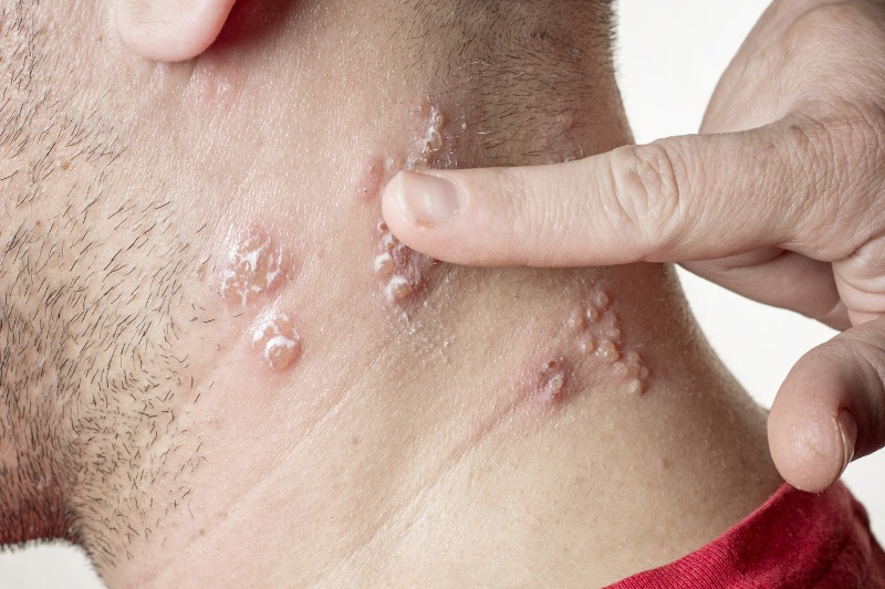


Penile cancer is a rare but clinically devastating malignant neoplasm of the male external genitalia, accounting for less than 1% of male malignancies in developed countries, though its incidence remains significantly higher in developing regions. Originating predominantly from the squamous epithelial lining of the glans penis, coronal sulcus, or inner prepuce, penile carcinoma presents a diagnostic and therapeutic challenge that balances strict oncological control with functional and psychosexual organ preservation. Early clinical recognition of subtle mucosal lesions, coupled with precise histopathological staging and regional lymph node assessment, is paramount to optimizing disease-free survival and patient quality of life.
1. Disease Overview and Pathophysiological Mechanisms
What is Penile Cancer?
Penile cancer is defined as an invasive malignant transformation of the cutaneous and mucosal epithelium of the penis. More than 95% of primary penile malignancies are classified histopathologically as Squamous Cell Carcinoma (SCC). Rare non-squamous subtypes include basal cell carcinoma, melanoma, sarcoma, and primary penile lymphoma.
Dual Etiological Pathways of Penile Carcinogenesis
Current oncological evidence establishes two distinct molecular and cellular pathways driving penile squamous cell carcinoma:
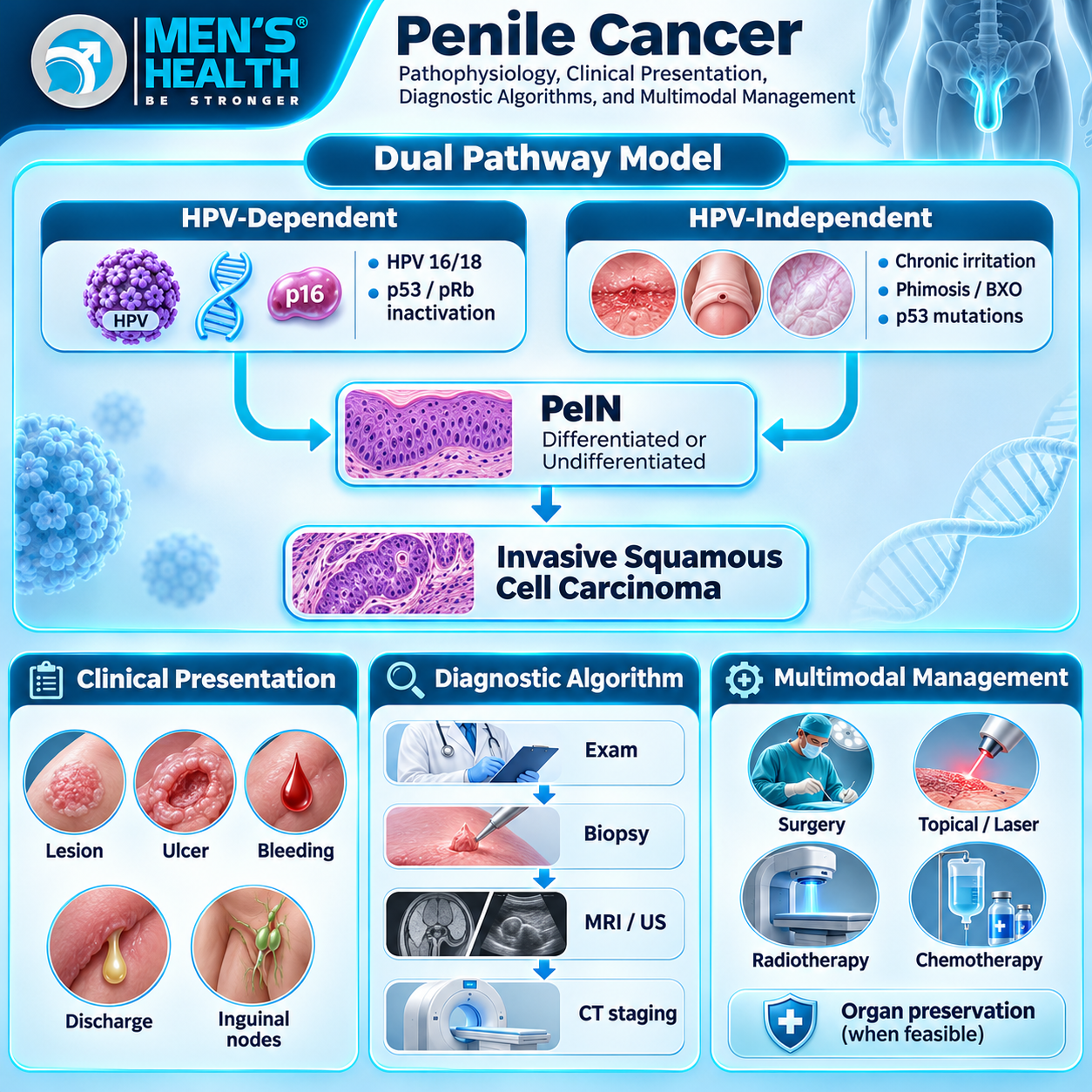
- Human Papillomavirus (HPV)-Dependent Pathway: Responsible for approximately 40% to 50% of cases. High-risk oncogenic genotypes (predominantly HPV-16 and HPV-18) integrate viral DNA into host keratinocytes. Expression of viral oncoproteins E6 and E7 leads to ubiquitin-mediated degradation of the tumor suppressor protein p53 and functional inactivation of the retinoblastoma protein (pRb), respectively, driving uncontrolled cellular proliferation and p16INK4a overexpression.
- HPV-Independent Pathway: Accounts for 50% to 60% of cases. This pathway is driven by chronic mechanical irritation, chronic inflammation, and persistent smegma accumulation secondary to severe phimosis. Over time, persistent inflammatory stress induces somatic mutations in the TP53 gene and dysregulates epidermal growth factor receptor (EGFR) signaling pathways, independent of viral integration.
2. Risk Factors and Premalignant Lesions
Understanding the epidemiological and anatomical risk factors facilitates early targeted clinical screening.
Primary Predisposing Factors
- Phimosis: The inability to retract the prepuce is present in 45% to 85% of patients with penile SCC. Phimosis impedes proper hygiene, leading to the accumulation of smegma, desquamated cells, and bacterial flora, inducing chronic balanoposthitis.
- Lack of Neonatal Circumcision: Neonatal circumcision offers strong protective efficacy against HPV-independent penile cancer by eliminating the preputial space and preventing chronic smegma retention. However, adult circumcision provides less protective benefit.
- High-Risk HPV Infection and Sexual Behaviors: Multiple sexual partners and a history of condyloma acuminata elevate lifetime exposure to oncogenic HPV strains.
- Tobacco Use: Cigarette smoking exerts a synergistic carcinogenic effect, with tobacco metabolites concentrates in genital secretions, increasing the risk of penile carcinoma 3- to 5-fold.
Premalignant Lesions (Penile Intraepithelial Neoplasia – PeIN)
Penile dysplasia is categorized histologically as PeIN (grade 1 to 3) and clinically manifests as:
- Erythroplasia of Queyrat: Non-keratinized, velvety, bright red plaque presenting on the glans or inner prepuce (HPV-associated).
- Bowen’s Disease: Solitary, well-demarcated, scaly, erythematous plaque located on the cutaneous shaft (HPV-associated).
- Lichen Sclerosus (Balanitis Xerotica Obliterans – BXO): Chronic autoimmune inflammatory dermatosis causing whitish, sclerotic plaques on the glans and meatus, strongly linked to HPV-independent differentiated PeIN.
3. Clinical Presentation and Physical Signs
Early detection relies on thorough physical inspection of the distal penis. In patients with uncorrected phimosis, lesions may remain concealed under an unretractable foreskin for months.
Primary Local Signs
- Focal Mucosal or Cutaneous Lesion: Ranges from a subtle indurated plaque, superficial exophytic warty growth, hyperkeratotic papule, or non-healing shallow ulceration on the glans (60%), prepuce (25%), or coronal sulcus (10%).
- Chronic Serosanguinous or Purulent Discharge: Unexplained malodorous discharge draining from beneath a phimotic prepuce, often accompanied by localized bleeding upon palpation.
- Palpable Inguinal Lymphadenopathy: Up to 50% of patients present with enlarged inguinal lymph nodes at initial diagnosis. While lymphadenopathy may reflect reactive inflammation secondary to secondary ulcer infection, 50% of palpable nodes harbour metastatic disease.
Red Flag Symptoms Requiring Urgent Urological Referral
- A non-healing ulcer or indurated nodule on the glans or prepuce persisting for >2 weeks.
- Spontaneous, recurrent bleeding from the preputial orifice.
- Hard, fixed, or matted bilateral inguinal nodal masses.
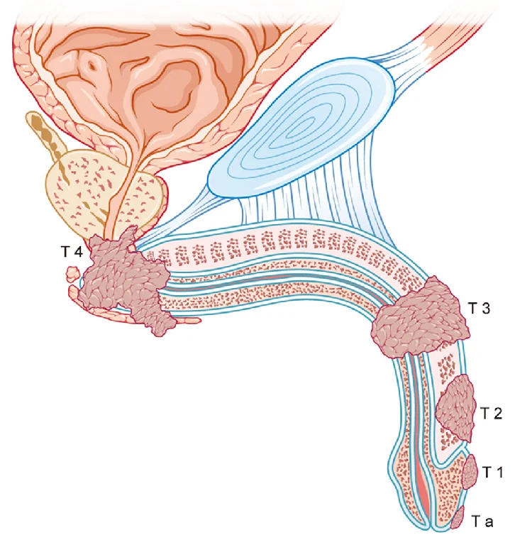
- Acute urinary retention or urethral fistula formation due to direct tumor invasion into the corpus spongiosum and urethral lumen (T3 disease).
4. Differential Diagnosis Spectrum
Penile lesions present a broad differential diagnosis spanning infectious, inflammatory, and benign neoplastic disorders.
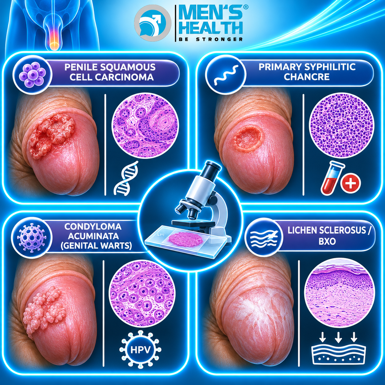
Diagnostic Comparison Matrix
| Condition | Physical Appearance | Palpation & Margins | Natural History | Diagnostic Standard |
| Penile SCC | Exophytic nodule or ulcerative defect | Hard, indurated base; irregular rolled edges | Progressive tissue invasion and destruction | Histopathological punch/excisional biopsy |
| Syphilitic Chancre | Single shallow, clean-based ulcer | Firm, rubbery, indurated, painless | Self-limiting (resolves in weeks without treatment) | Darkfield microscopy, Treponemal serology |
| Condyloma Acuminata | Soft, cauliflower-like papules | Non-indurated, flexible base | Benign growth, frequent local recurrence | Clinical exam, viral typing |
| Lichen Sclerosus | White sclerotic plaques, scarring | Atrophic, loss of mucosal elasticity | Chronic progressive autoimmune inflammation | Clinical exam, target biopsy |
| Peyronie’s Disease | Subcutaneous fibrous plaque | Dense plaque on shaft, no skin ulceration | Causes penile curvature during erection | Clinical exam, Penile Doppler US |
5. Diagnostic Pathways and Staging Protocols
Definitive evaluation of penile cancer requires histopathological confirmation of the primary lesion combined with systematic assessment of regional lymph nodes and distant organ sites.
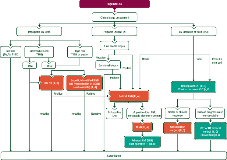
Diagnostic Workflow
- Primary Tumor Biopsy: Punch biopsy or full-thickness incisional biopsy of the lesion edge is mandatory to establish histological subtype, depth of invasion, microvascular invasion, and histological grade (G1 to G3).
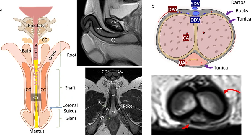
- Local Imaging: High-resolution Magnetic Resonance Imaging (MRI) of the penis (with artificial erection induced by intracavernosal prostaglandin E1 injection) evaluates tumor infiltration into the tunica albuginea, corpora cavernosa, and urethral spongiosum (differentiating T1/T2 from T3 disease).
- Inguinal Lymph Node Staging:
- In cN0 (Non-palpable nodes): High-risk features (Stage ≥ T1b, high grade, lymphovascular invasion) warrant Dynamic Sentinel Lymph Node Biopsy (DSLNB) or bilateral superficial modified inguinal lymphadenectomy.
- In cN1/N2 (Palpable nodes): Ultrasound-guided Fine Needle Aspiration Cytology (FNAC) or core biopsy confirms metastatic spread.
- Distant Staging: Contrast-enhanced Chest, Abdomen, and Pelvic CT, or 18F-FDG PET-CT, is indicated in node-positive disease to detect pelvic nodal metastasis or visceral organ involvement.
6. Treatment Framework: Surgical and Systemic Modalities
Management of penile cancer is stratified based on the primary tumor stage (T-stage), regional nodal status (N-stage), and distant metastasis (M-stage).
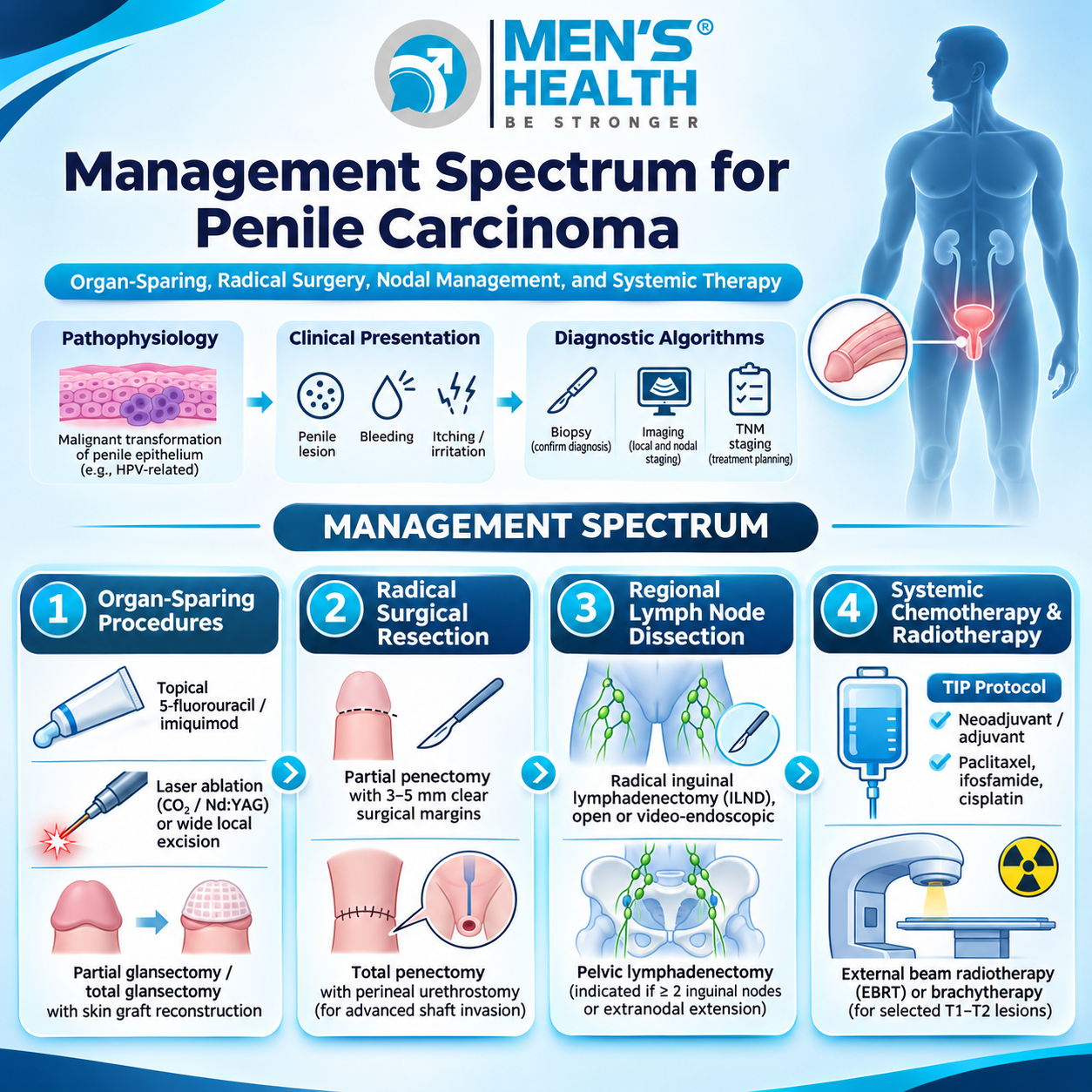
1. Primary Tumor Management
- Organ-Sparing Surgery: Modern urological guidelines prioritize penile preservation whenever oncologically safe. For surface dysplasia (PeIN) or small localized T1 lesions, resurfacing of the glans, laser ablation, or partial glansectomy offers excellent local control while preserving sexual and urinary function.
- Amputative Surgery: Invasive tumors infiltrating deep cavernosal structures (T2–T4) require partial penectomy with a 3- to 5-mm negative surgical margin, or total penectomy with perineal urethrostomy creation when shaft length is insufficient for standing micturition.
2. Regional Lymph Node Management
Inguinal lymph node control is the strongest determinant of long-term survival:
- Radical Inguinal Lymphadenectomy (ILND): Removal of superficial and deep inguinal nodal tissue.
- Pelvic Lymphadenectomy: Performed when two or more inguinal nodes show metastasis or when Extranodal Extension (ENE) is identified.
3. Systemic Pharmacotherapy
Systemic multi-agent chemotherapy plays a vital role in advanced and node-positive disease:
- Neoadjuvant Chemotherapy (TIP Protocol): Paclitaxel, Ifosfamide, and Cisplatin administered prior to radical ILND in patients with fixed, bulky, or ulcerated inguinal nodes to downstage disease.
- Adjuvant Chemotherapy: Recommended following nodal resection when multiple nodes or extranodal extension are present.
7. Pharmacological Safety, Side Effects, and Drug Interactions
Systemic oncological regimens for penile cancer involve potent chemotherapeutic agents requiring strict clinical monitoring.
- Cisplatin Toxicity: Induces significant nephrotoxicity, ototoxicity, and severe emesis. Vigorous intravenous hydration with isotonic saline before and after infusion is mandatory. Concomitant use of aminoglycosides or loop diuretics exacerbates renal and auditory damage.
- Ifosfamide Toxicity: Associated with hemorrhagic cystitis due to the metabolite acrolein. Concomitant administration of Mesna (2-mercaptoethane sulfonate sodium) and hyperhydration is mandatory to prevent urothelial toxicity. Encephalopathy (ifosfamide-induced CNS toxicity) requires monitoring.
- Paclitaxel Reactions: Peripheral neuropathy and hypersensitivity reactions. Pre-medication with corticosteroids, H1-antagonists, and H2-antagonists is required. Hepatic CYP2C8 and CYP3A4 inhibitors (e.g., ketoconazole, clarithromycin) increase paclitaxel exposure and toxicity.
8. Role of Men’s Health Center and Men’s Health Pharmacy
Navigating the complex diagnostic, surgical, and psychological pathways of male genital malignancies requires a highly specialized, multidisciplinary medical network.
The Nhà thuốc Nam khoa Men’s Health (Men’s Health Andrology Pharmacy), operating within the ecosystem of the Trung tâm Sức khỏe Nam giới Men’s Health (Men’s Health Center) founded by TS.BS.CK2 Trà Anh Duy (Dr. Tra Anh Duy, MD, PhD, Spec. II), offers clinical pharmacy support for male urological and oncological care. At Men’s Health, patients consult with experienced urologists and clinical pharmacists who provide clear guidance on pre-operative prep, post-operative wound care, systemic medication management, and symptom relief. The center emphasizes professional standards, patient safety, discreet consultation, strict information confidentiality, and personalized care throughout diagnostic evaluation, surgery, and post-treatment follow-up. All prescription medications and specialized therapies are managed under direct medical supervision, highlighting the collaborative effort between physicians and clinical pharmacists to ensure optimal therapeutic efficacy and patient safety.

9. Lifestyle Modifications, Prevention, and Common Errors
Primary Prevention Strategies
- HPV Vaccination: Administering the 9-valent HPV vaccine to young males prior to sexual debut significantly reduces the risk of HPV-associated PeIN and penile SCC.
- Correction of Phimosis: Early surgical management of phimosis restores penile hygiene and eliminates chronic preputial irritation.
- Rigorous Genital Hygiene: Regular retraction and thorough cleaning of the preputial sac prevent smegma accumulation.
- Smoking Cessation: Eliminates synergistic carcinogenic exposures.
Common Clinical Errors to Avoid
- Delaying Medical Evaluation Due to Stigma: Patients frequently defer seeking care for 6 to 12 months out of embarrassment, allowing localized disease to progress to advanced stages requiring radical amputation.
- Empiric Prescription of Topical Steroids or Antibiotics Without Biopsy: Treating a chronic, non-healing penile ulcer with topical antifungal or steroid creams for months without performing a tissue biopsy delays definitive diagnosis.
- Omitting Inguinal Node Evaluation: Treating the primary penile tumor while neglecting regional lymph node staging, leading to unmanaged regional metastatic spread.
Penile Cancer: Frequently Asked Questions
What are the earliest visual signs of penile cancer?
The earliest signs include a small, persistent, red velvety patch, an indurated skin nodule, a shallow painless sore, or a warty growth on the glans or inner foreskin that does not heal within two weeks.
Is penile cancer contagious to a sexual partner?
No. Penile cancer itself is not contagious. However, the Human Papillomavirus (HPV), which contributes to a significant portion of penile cancers, is a sexually transmitted infection. HPV vaccination and barrier contraception reduce viral transmission.
Does treating penile cancer always require complete removal of the penis?
No. Modern urological protocols emphasize organ-sparing techniques (such as laser ablation, glans resurfacing, or partial glansectomy) for early-stage (Tis, T1) lesions, preserving urinary function and sexual potency whenever oncologically safe.
How does phimosis increase the risk of penile cancer?
Phimosis prevents foreskin retraction, leading to the accumulation of smegma, dead skin cells, and bacteria. This causes chronic recurrent balanoposthitis, which induces long-term inflammatory stress and cellular dysplasia in the mucosal epithelium.
Can penile cancer be cured if detected early?
Yes. Early-stage penile cancer localized to the glans or prepuce without lymph node involvement achieves 5-year survival rates exceeding 85% to 90% following appropriate surgical resection and staging.
Conclusion and Medical Advice
Penile cancer is a serious but highly treatable malignancy when detected during its early localized stages. Recognizing cardinal clinical features – such as persistent mucosal plaques, non-healing glans ulcers, or unexplained preputial discharge – enables prompt biopsy and accurate histopathological staging. Organ-sparing surgical techniques, coupled with rigorous inguinal lymph node evaluation and systemic chemotherapy when indicated, maximize oncological cure while preserving male anatomical and functional integrity.
Individuals who notice an persistent lesion, skin change, or abnormal discharge beneath the foreskin should avoid self-medication, topical home remedies, or delayed observation due to embarrassment. Seeking timely evaluation at a specialized center such as the Trung tâm Sức khỏe Nam giới Men’s Health guarantees diagnostic precision, expert surgical management, and dedicated clinical pharmacy support within a confidential, professional, and supportive care environment.