

Testicular cancer represents the most frequent solid malignancy affecting young male adults aged 15 to 44 years. Characterized predominantly by Testicular Germ Cell Tumors (TGCTs), this malignancy develops via malignant transformation of primordial germ cells within the seminiferous tubules. Despite its malignant potential, testicular cancer exhibits extraordinary sensitivity to platinum-based chemotherapy regimens and surgical resection, achieving long-term disease-free survival rates exceeding 95% when detected early. Understanding the underlying etiology, recognizing cardinal diagnostic signs, establishing differential diagnoses, and utilizing multi-modal staging protocols are essential for preserving both oncological safety and male reproductive potential.
1. Clinical Overview and Embryological Pathophysiology
What is Testicular Cancer?
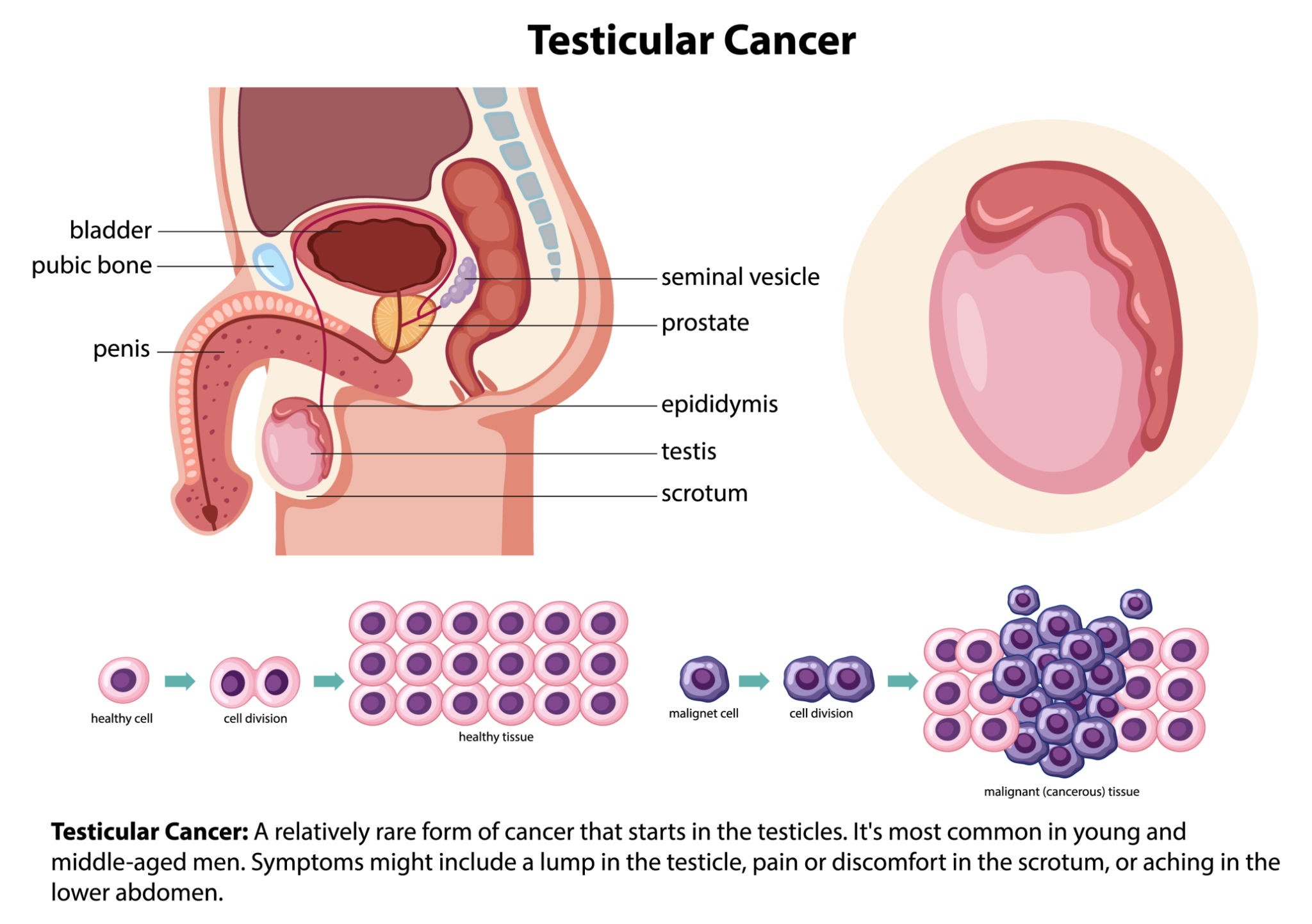
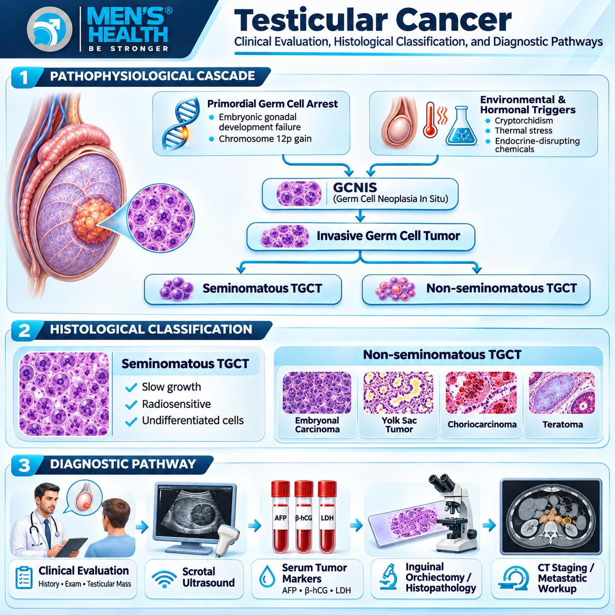
Testicular cancer is a malignant neoplasm originating from the parenchymal cellular components of the testis – most commonly the germinal epithelium responsible for spermatogenesis. More than 95% of primary testicular tumors are Testicular Germ Cell Tumors (TGCTs), which derive from a precursor lesion termed Germ Cell Neoplasia In Situ (GCNIS).
Histological Classification
TGCTs are divided into two main clinical and histological categories, which differ significantly in management and prognosis:
- Seminomatous Germ Cell Tumors (Seminomas): Comprising approximately 50% of TGCTs, seminomas consist of uniform, undifferentiated primordial germ cells. They tend to grow relatively slowly, metastasize late via predictable lymphatic routes to retroperitoneal lymph nodes, and demonstrate high sensitivity to both radiotherapy and platinum-based chemotherapy.
- Non-Seminomatous Germ Cell Tumors (NSGCTs): Comprising embryonal carcinoma, yolk sac tumor, choriocarcinoma, and teratoma (often occurring as mixed germ cell tumors). NSGCTs are characterized by pluripotent differentiation, rapid cellular kinetics, early hematogenous vascular dissemination, and resistance to radiotherapy.
- Non-Germ Cell Tumors (Sex Cord-Stromal Tumors): Account for 3% to 5% of testicular neoplasms, including Leydig cell tumors and Sertoli cell tumors. These neoplasms frequently secrete functional steroid hormones (estrogens or androgens), manifesting clinically with gynecomastia, precocious puberty, or endocrine dysfunction.
2. Etiology and Risk Factor Profile
While the exact molecular initiators of testicular cancer remain a subject of active research, several established congenital, genetic, and environmental risk factors elevate individual susceptibility.
Primary Predisposing Risk Factors
- Cryptorchidism (Undescended Testis): The single strongest clinical risk factor. Males born with cryptorchidism face a 4- to 10-fold increased risk of developing testicular cancer in the affected testis, as well as a slightly elevated risk in the contralateral scrotum. The abnormal higher temperature of the inguinal canal or retroperitoneum disrupts germ cell maturation and promotes malignant transformation.
- Personal or Family History of TGCT: A previous history of testicular cancer in one testis increases the risk of developing a metachronous primary tumor in the contralateral testis by 12- to 18-fold. First-degree relatives (brothers or fathers) of affected individuals exhibit a 4- to 8-fold risk elevation, highlighting hereditary genetic susceptibility (e.g., variants in KITLG, SPRY4, and BAK1).
- Testicular Dysgenesis Syndrome (TDS): A developmental disorder originating in embryonic life, encompassing cryptorchidism, hypospadias, impaired spermatogenesis (subfertility), and GCNIS development, driven by disrupted fetal Sertoli/Leydig cell differentiation.
- Environmental and Industrial Exposures: Maternal or early-life exposure to endocrine-disrupting chemicals (EDCs), including organochlorine pesticides, phthalates, and synthetic estrogens.
3. Clinical Manifestations and Red Flag Symptoms
Early recognition of cardinal physical signs is the cornerstone of prompt diagnosis. Patients frequently discover abnormalities during routine personal hygiene or partner contact.
Common Signs and Symptoms
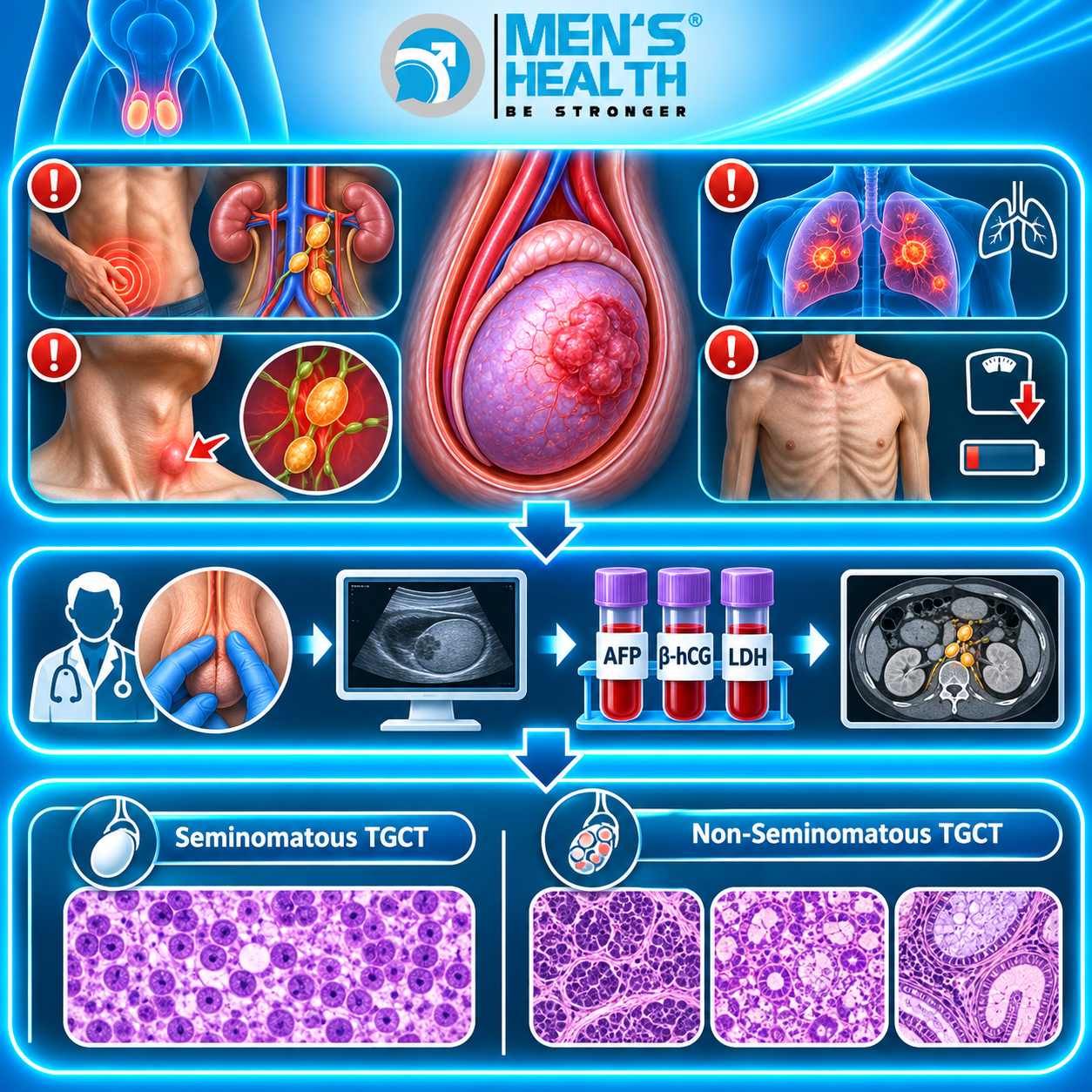
- Painless Testicular Nodule or Enlargement: The classic presentation (present in >80% of cases) is a firm, painless, non-tender nodule or diffuse induration originating within the testicular parenchyma.
- Scrotal Heaviness or Dull Pelvic Ache: A sensation of dragging, fullness, or persistent dull discomfort in the lower abdomen, groin, or scrotum.
- Acute Scrotal Pain (10%–15% of cases): Occurs when rapidly growing tumors undergo intra-tumoral hemorrhage, infarction, or secondary necrosis, mimicking acute epididymitis or testicular torsion.
- Endocrine Symptoms: Bilateral gynecomastia (breast tenderness or enlargement) resulting from human Chorionic Gonadotropin (hCG) secretion by syncytiotrophoblastic cells, or systemic hyperestrogenism from sex cord-stromal tumors.
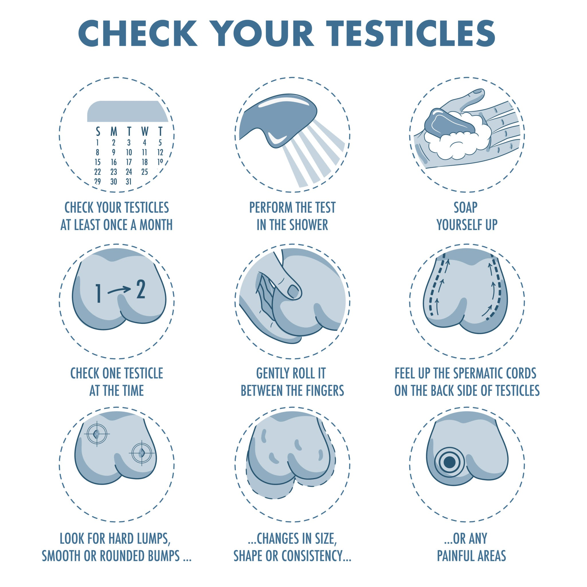
Diagnostic Physical Examination & Testicular Self-Examination (TSE)
Routine monthly Testicular Self-Examination (TSE) allows men to familiarize themselves with normal testicular architecture, facilitating early detection of focal indurations.
4. Differential Diagnosis Spectrum
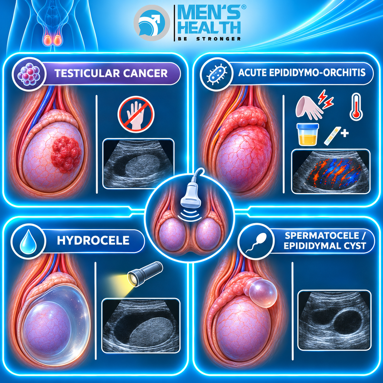
Many non-malignant intrascrotal conditions share clinical features with testicular cancer. Accurately differentiating malignant parenchymal masses from extra-testicular fluid collections or infectious lesions is essential to avoid unnecessary radical surgery or delayed oncological therapy.
Diagnostic Comparison Matrix
| Clinical Condition | Primary Anatomical Location | Physical Exam & Palpation | Transillumination | Doppler Ultrasound Findings |
| Testicular Cancer | Intratesticular parenchyma | Firm, hard, non-tender nodule | Negative (Opaque mass) | Focal hypoechoic lesion, hypervascular flow |
| Epididymo-Orchitis | Epididymis & Testis | Severely tender, warm, swollen | Negative | Diffuse parenchymal hyperemia, enlarged epididymis |
| Hydrocele | Tunica vaginalis space | Fluid-filled, soft, non-tender | Positive (Transilluminates) | Anechoic fluid surrounding normal testis |
| Spermatocele | Head of epididymis | Smooth, mobile mass above testis | Positive | Well-circumscribed, anechoic cyst separate from testis |
| Testicular Torsion | Spermatic cord / Testis | Exquisitely tender, high-riding | Negative | Absent or severely reduced arterial flow |
5. Comprehensive Clinical Diagnostic Pathways
Establishing an accurate diagnosis requires a structured pathway moving from non-invasive screening and biomarker profiling to definitive histopathological confirmation.
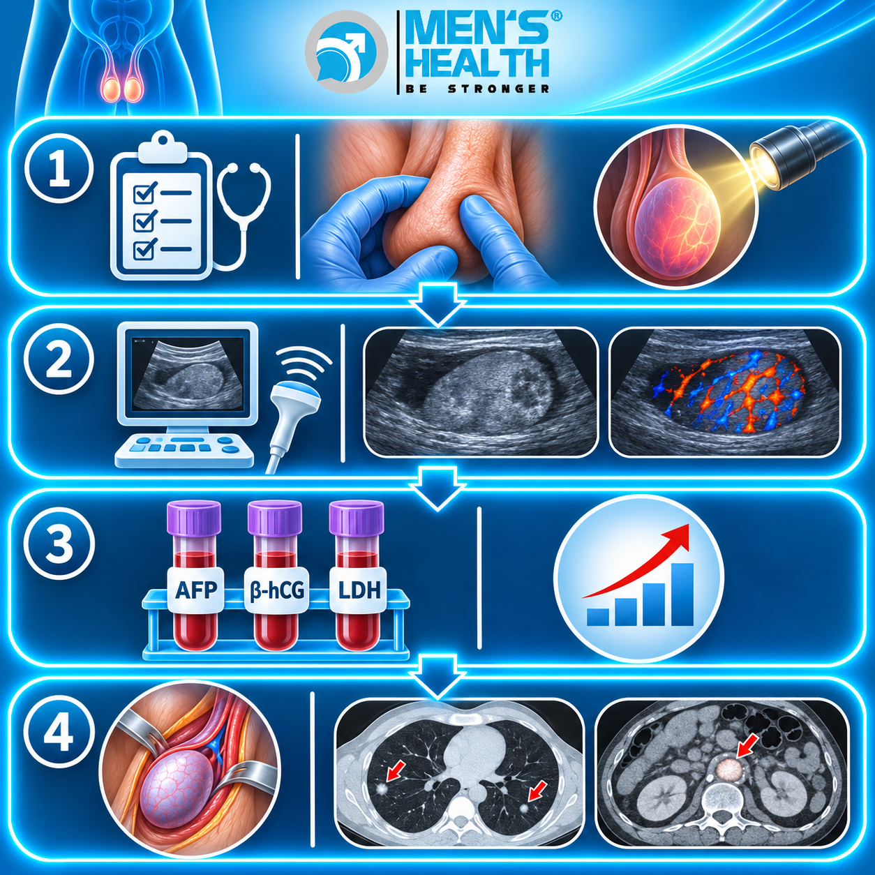
- High-Resolution Scrotal Ultrasonography: The non-invasive imaging modality of choice (>99% diagnostic sensitivity). High-frequency ultrasound differentiates intratesticular lesions from extratesticular pathology. Malignant tumors typically appear as focal, hypoechoic parenchymal masses with internal vascular signals on Color Doppler.
- Serum Tumor Markers (STMs): Pre-operative baseline measurement of three serum proteins provides vital diagnostic, histological, and prognostic information:
- Alpha-Fetoprotein (AFP): Produced by yolk sac cells and embryonal carcinoma. Elevated serum AFP rules out pure seminoma regardless of histological appearance.
- Beta-Human Chorionic Gonadotropin (β-hCG): Secreted by syncytiotrophoblastic cells. Markedly elevated in choriocarcinoma and moderately elevated in 15% to 20% of seminomas.
- Lactate Dehydrogenase (LDH): A non-specific enzyme reflecting tumor burden, cellular turnover, and tissue breakdown.
- Radical Inguinal Orchiectomy: Trans-scrotal biopsy or aspiration is strictly contraindicated due to the high risk of tumor seeding into superficial scrotal lymphatics. Definitive tissue diagnosis requires radical inguinal orchiectomy – resecting the testis, epididymis, and spermatic cord up to the internal inguinal ring via an inguinal incision.
- Staging Modalities: Post-orchiectomy staging includes contrast-enhanced Computed Tomography (CT) scans of the chest, abdomen, and pelvis to evaluate retroperitoneal lymph nodes and visceral organ involvement.
6. Treatment Protocols and Systemic Management
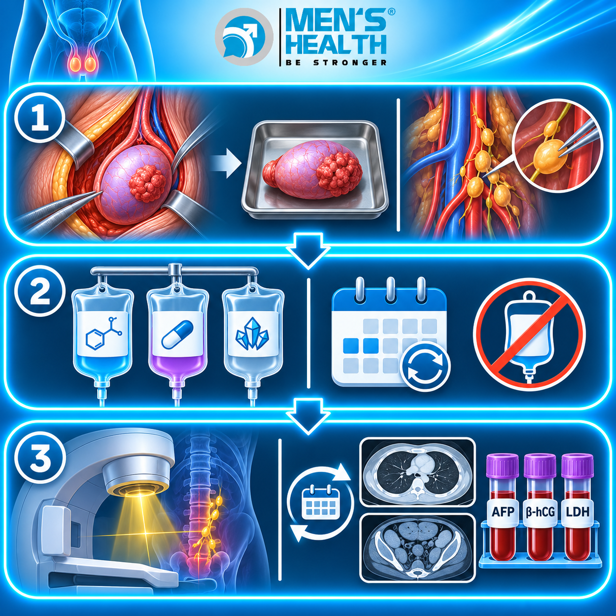
Management strategies for testicular cancer depend on histological subtype (seminoma vs. non-seminoma), clinical stage (Stage I, II, or III), and IGCCCG (International Germ Cell Cancer Collaborative Group) risk stratification.
Primary Pharmacotherapy Regimens
- BEP Protocol (Bleomycin, Etoposide, Cisplatin): The international standard multi-agent chemotherapy regimen for advanced or metastatic TGCTs. Administered in 3 or 4 cycles depending on risk classification.
- EP Protocol (Etoposide, Cisplatin): Utilized for patients at risk of pulmonary toxicity from Bleomycin or elderly individuals with compromised renal reserve.
7. Side Effects, Contraindications, and Pharmacological Safety
Chemotherapeutic protocols for testicular cancer are highly effective but carry specific toxicities that require close medical monitoring:
- Cisplatin Toxicity: Nephrotoxicity, severe nausea, peripheral neuropathy, and irreversible high-frequency ototoxicity. Requires intensive pre- and post-hydration with isotonic saline.
- Bleomycin Pulmonary Toxicity: Pulmonary fibrosis and pneumonitis. Serial pulmonary function tests (DLCO) are mandatory; Bleomycin must be discontinued immediately if lung diffusion capacity declines significantly.
- Etoposide Myelosuppression: Grade 3–4 neutropenia, anemia, and thrombocytopenia, elevating the risk of neutropenic fever. Secondary leukemogenesis (acute myeloid leukemia) represents a rare long-term risk.
8. Fertility Preservation Protocols and Reproductive Care
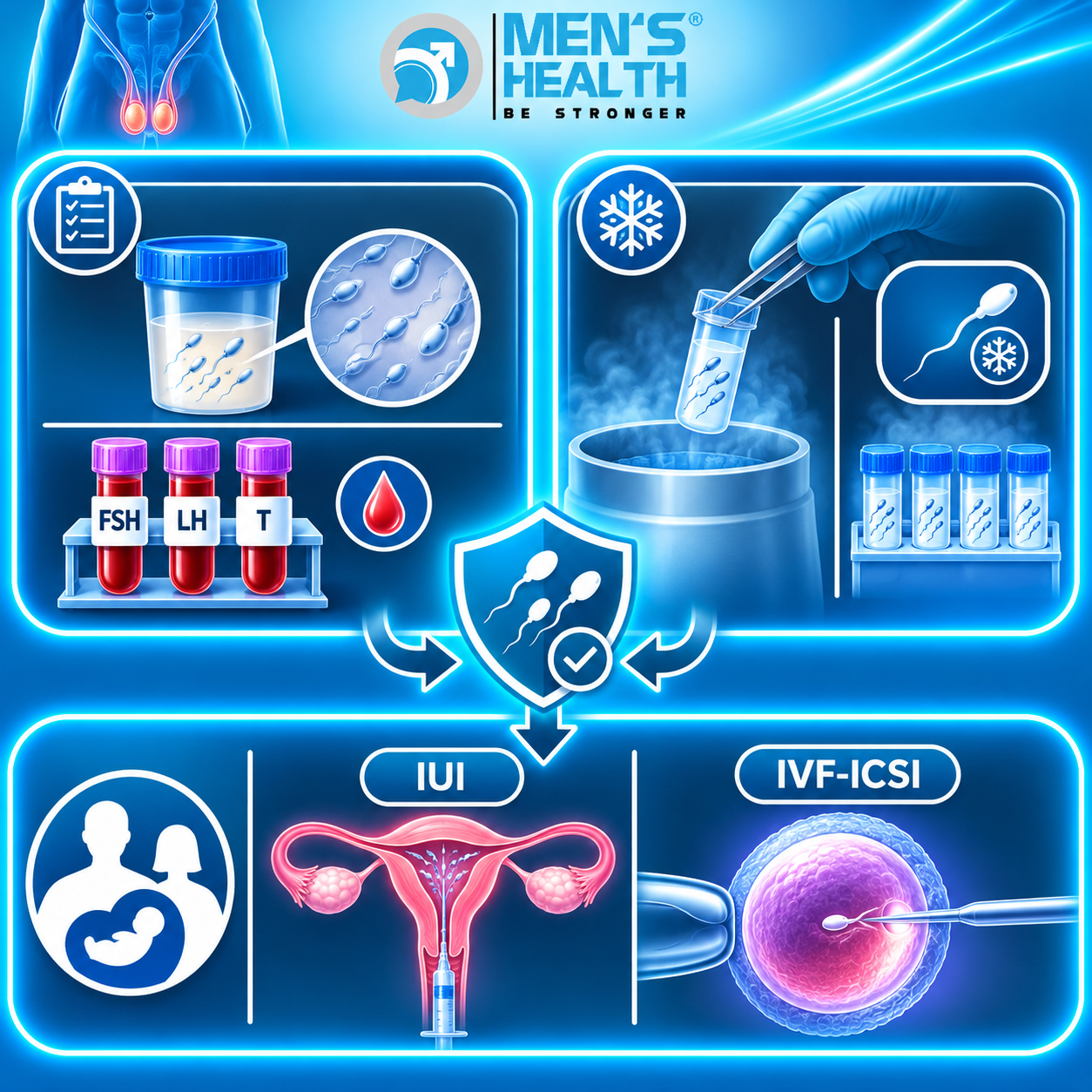
Because testicular cancer disproportionately affects young men in their reproductive years, discussing fertility preservation prior to initiating definitive treatment is essential.
Reproductive Preservation Measures
- Pre-Treatment Sperm Cryopreservation: Patients should undergo baseline semen analysis and sperm cryopreservation before radical inguinal orchiectomy, systemic chemotherapy, or retroperitoneal lymph node dissection (RPLND).
- Impact of Chemotherapy and Radiation: Systemic chemotherapy and pelvic radiation induce transient or permanent azoospermia and elevated sperm DNA fragmentation. Recovery of spermatogenesis can take 2 to 5 years post-treatment.
- Ejaculatory Dysfunction Post-RPLND: Nerve-sparing RPLND techniques help preserve sympathetic post-ganglionic fibers responsible for seminal emission, preventing retrograde ejaculation and anejaculation.
9. Professional Care at Men’s Health Center and Men’s Health Pharmacy
Navigating testicular cancer screening, diagnostic evaluation, surgical coordination, and fertility preservation requires dedicated medical expertise and multidisciplinary pharmaceutical support.
Nhà thuốc Nam khoa Men’s Health (Men’s Health Andrology Pharmacy), an integral unit within the Trung tâm Sức khỏe Nam giới Men’s Health (Men’s Health Center) ecosystem founded by TS.BS.CK2 Trà Anh Duy (Dr. Tra Anh Duy, MD, PhD, Spec. II), provides dedicated clinical pharmacy support for male reproductive and urological health. At Men’s Health, patients consult with experienced urologists and clinical pharmacists who provide personalized guidance on medication safety, supportive care management, and long-term reproductive tracking. The facility maintains strict clinical safety, privacy, absolute confidentiality, and evidence-based practice across all stages of care. Prescription medications and post-operative therapeutic regimens are dispensed strictly under physician supervision, emphasizing the essential collaborative role between physicians and clinical pharmacists in ensuring optimal patient safety and therapeutic outcomes.

10. Common Diagnostic Pitfalls and Clinical Errors
- Delaying Evaluation of a Painless Testicular Nodule: Dismissing a firm, painless intratesticular mass as a minor injury or benign cyst, leading to delayed diagnosis and higher disease stage.
- Performing Trans-Scrotal Biopsy or Aspiration: Violating the scrotal barrier via trans-scrotal needle aspiration or biopsy, which alters lymphatic drainage and risks local tumor seeding into superficial inguinal lymph nodes.
- Misdiagnosing Testicular Cancer as Acute Epididymitis: Misinterpreting tumor-associated scrotal discomfort as an infection and prescribing repeated courses of oral antibiotics without obtaining a scrotal ultrasound.
- Omitting Pre-Treatment Sperm Cryopreservation: Initiating radical surgery, chemotherapy, or radiotherapy without offering sperm banking to young men desiring future biological children.
Testicular Cancer: Frequently Asked Questions
What are the earliest warning signs of testicular cancer?
The classic early sign is a firm, painless nodule, lump, or swelling within the body of the testis. Some men experience a heavy dragging sensation in the scrotum or a dull ache in the lower abdomen or groin.
Is testicular cancer curable?
Yes. Testicular cancer is one of the most curable solid malignancies, achieving overall survival rates exceeding 95%. When detected in early, localized stages (Stage I), cure rates approach 99%.
Why is trans-scrotal needle biopsy forbidden in suspected testicular cancer?
The scrotal skin drains to superficial inguinal lymph nodes, whereas the testes drain to deep retroperitoneal lymph nodes. Performing a needle biopsy through the scrotum breaks anatomical barriers and increases the risk of spreading tumor cells to the inguinal lymphatic basin.
Does removing one testicle cause permanent infertility or low testosterone?
Unilateral orchiectomy leaves one healthy contralateral testicle, which undergoes compensatory hypertrophy to produce sufficient testosterone and sperm for normal fertility and sexual function. However, because baseline fertility may already be altered in cryptorchid men, pre-operative sperm cryopreservation is strongly recommended.
How often should men perform testicular self-examination (TSE)?
Men aged 15 to 44 – especially those with a history of cryptorchidism or a family history of testicular cancer – should perform a gentle monthly testicular self-examination after a warm shower or bath.
Conclusion and Expert Medical Advice
Testicular cancer is a highly treatable malignancy when identified early through routine clinical evaluation, high-resolution scrotal ultrasonography, and serum tumor marker testing. Recognizing cardinal physical signs – such as a firm, painless intratesticular mass – and avoiding common diagnostic errors (such as trans-scrotal biopsy or delayed ultrasound evaluation) are critical for optimizing treatment success.
Men who detect a mass, nodule, or persistent structural change within the scrotum should avoid self-diagnosis or unverified treatments. Seeking immediate evaluation at a specialized center such as the Trung tâm Sức khỏe Nam giới Men’s Health guarantees accurate diagnostic assessment, expert surgical coordination, and professional clinical pharmacy support within a safe, professional, and confidential environment.